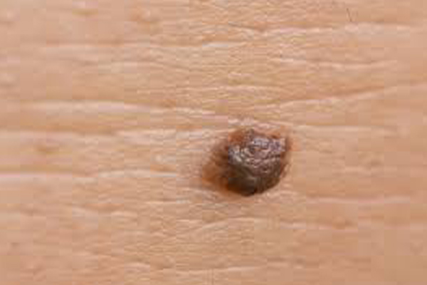
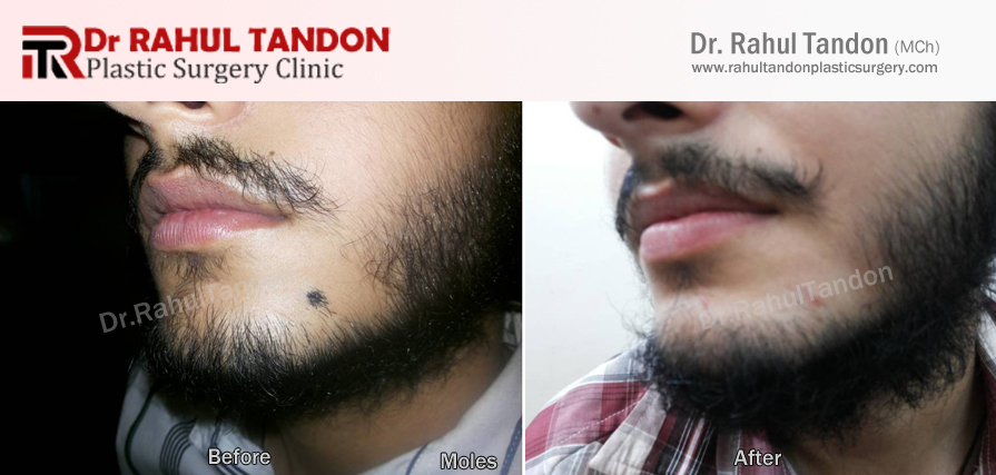
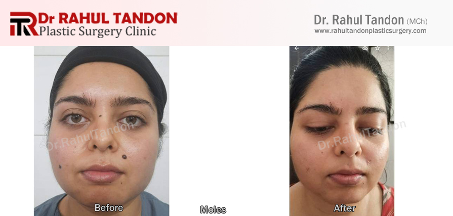
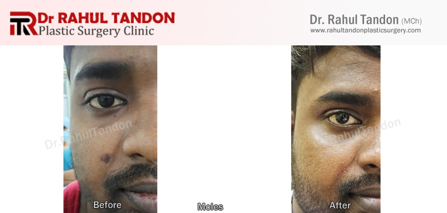
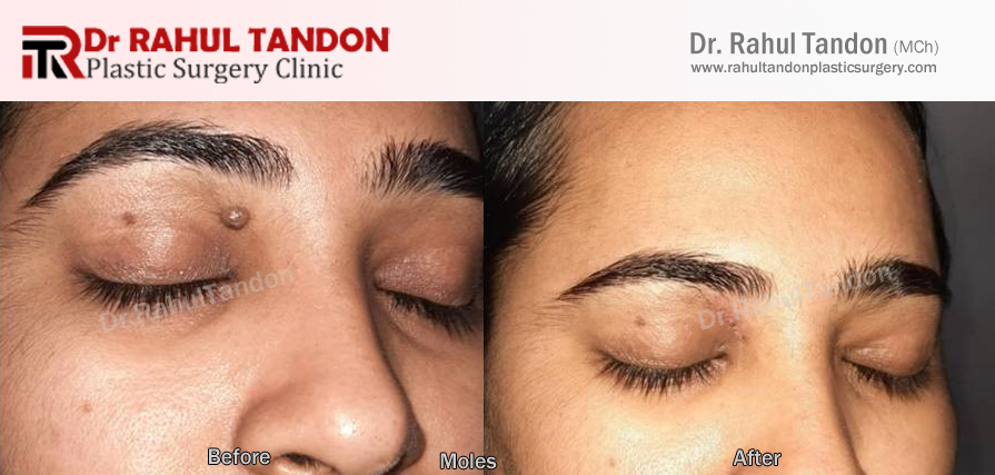
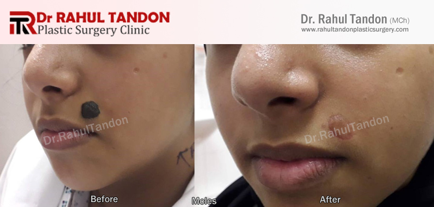
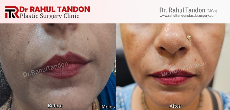
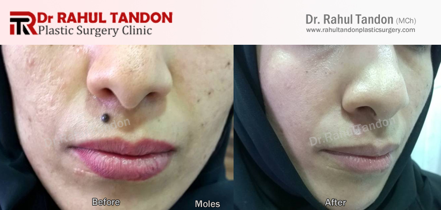
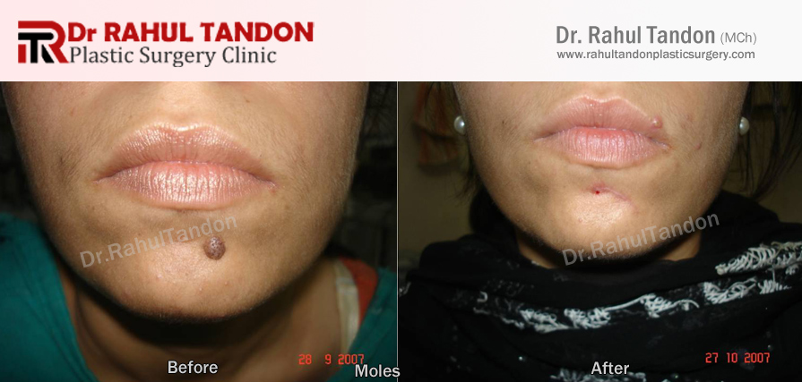
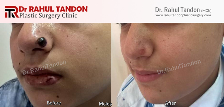
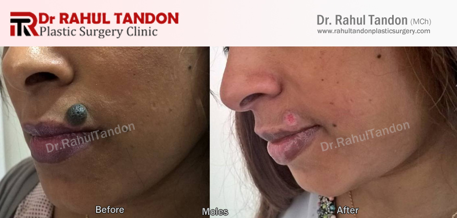
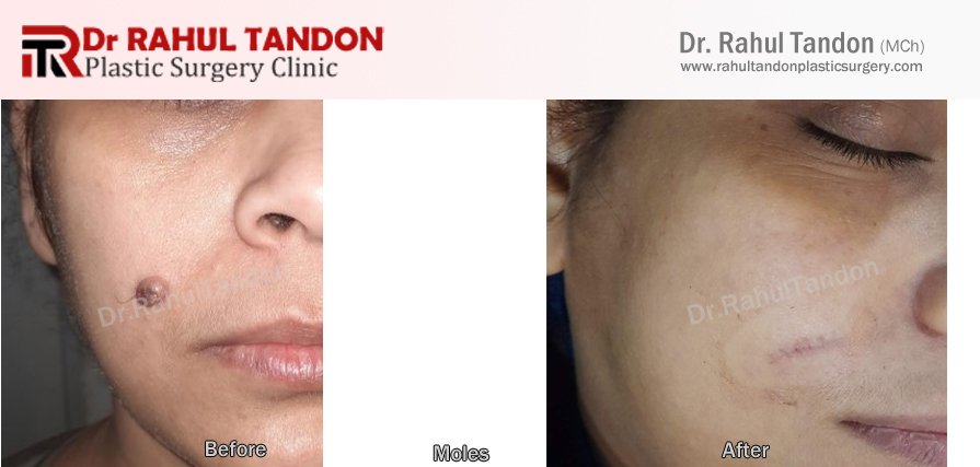
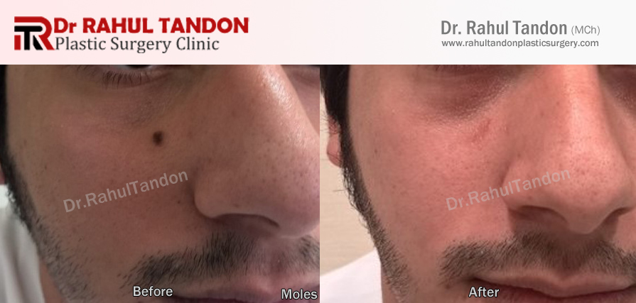
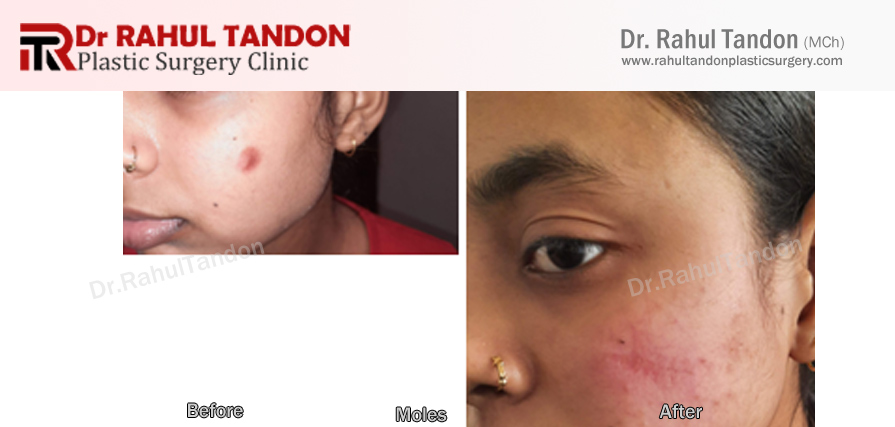
Moles are growths on the skin that are usually brown or black. Moles can appear anywhere on the skin, alone or in groups.
Most moles appear in early childhood and during the first 30 years of a person's life. It is normal to have between 10-40 moles by adulthood.
As the years pass, moles usually change slowly, becoming raised and/or changing color. Often, hairs develop on the mole. Some moles may not change at all, while others may slowly disappear over time.
One plans to get a mole removed if
- If he or she doesn’t like the position and appearance of mole over face or any other part of the body.
- If there has been an increase in size of the mole with or without appearance of hairs. The latter needs to be regularly trimmed which is quite disturbing for the patient.
TREATMENT
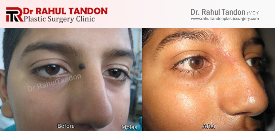
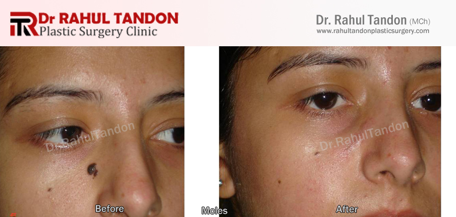
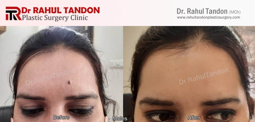
Moles can be removed on an outpatient basis under local anesthesia.
Smaller ones (< 3mm) or the ones which are located close to the vital parts of the face can be removed by laser or radiofrequency.
Bigger ones (> 3-4 mm) are removed by excision
There is minimal scarring after the procedure.
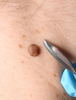
MELANOCYTIC NEVI
A nevus on the face, commonly known as a mole, is a benign growth of pigment-producing cells (melanocytes). While most facial nevi are harmless, some types carry a higher risk of developing into melanoma, a serious form of skin cancer. A mole's appearance can vary in color, size, and shape, and some may have hair growing from them.
Common types of facial nevi
- Intradermal nevus: A classic, raised, dome-shaped mole that is often skin-colored or light brown. The nevus cells are located in the deeper layer of the skin (dermis). They are very common on the face and neck.
- Compound nevus: This type has features of both a flat (junctional) and a raised (intradermal) mole. It is typically a brown, elevated bump surrounded by a flatter patch of pigment.
- Spitz nevus: A raised, dome-shaped mole that is usually pink but can be red, blue, or brown. Though benign, its appearance can sometimes be hard to distinguish from melanoma, especially in children and young adults. Spitz nevi often appear on the face.
- Nevus of Ota: A type of congenital dermal melanocytosis, it appears as a bluish-gray or brownish, patchy pigmentation on the face, typically on one side along the eye and temple. It is most common in individuals of Asian and African descent. It can be associated with an increased risk of glaucoma and, in rare cases, ocular or cutaneous melanoma, especially in Caucasian individuals.
- Nevus sebaceus: A congenital growth that most commonly affects the scalp but can also appear on the face. It looks like a solitary, smooth, yellowish plaque at birth. During puberty, it becomes more prominent and warty.
When to see a doctor
While most facial nevi are benign, you should see a dermatologist if you notice any changes. These signs can be remembered using the ABCDE guide:
- Asymmetry: One half of the mole does not match the other half.
- Border: The border is irregular, notched, or scalloped.
- Color: The color is uneven or includes multiple shades (tan, brown, black, blue, red, or white).
- Diameter: The mole is larger than 6 mm (about the size of a pencil eraser).
- Evolving: Any change in size, shape, color, or height, or the appearance of new symptoms like bleeding or itching.
Treatment options
Most benign facial nevi do not require treatment. However, they can be removed for cosmetic reasons or if they are suspicious for melanoma.
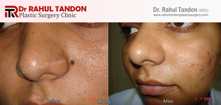
- Surgical excision: For suspicious lesions, the mole is surgically removed, and a pathologist examines the tissue for cancer cells.
- Shave excision: This involves using a razor to shave off the raised portion of the mole, which is suitable for moles that are clearly benign.
- Laser removal: Lasers can be used to treat some nevi, particularly blue nevi and nevus of Ota, especially when removal is for cosmetic purposes.
- Dermabrasion: This technique uses a wire brush to remove the top layers of skin, which can help lighten congenital nevi but may not remove all nevus cells.